
Research
The goal of the LAMPE (Laser-Assisted Medical Physics and Engineering) Lab is to develop optical methods based on Raman Spectroscopy (RS) and Non-Linear Optical (NLO) imaging including coherent Raman imaging, that enable rapid, non-invasive, cost-effective and highly accurate disease diagnostics and treatment. Using near-infrared laser light to excite vibrations of molecular chemical bonds (in RS), and nonlinear interactions with molecules (in NLO imaging), these methods provide highly detailed chemical, structural and functional information about cells and tissues in real time, without the use of external contrast agents (label-free). In combination with machine learning techniques, this enables us to effectively identify, quantify and differentiate between the response of cells and tissue exposed to different types of external stress such as disease and ionizing radiation. The ultimate goal of this research is to translate these optical techniques to the clinical and diagnostic lab setting.
Current Projects
Raman spectroscopy and machine learning for the identification of diseased and irradiated blood.
Current biochemical methods for the assessment of cellular and tissue response to stress inflicted by factors such as cancer and ionizing radiation require complex sample preparation and assess only a single stress biomarker. In contrast, Raman spectroscopy along with machine learning techniques enables rapid molecular fingerprinting of single cells, solid and liquid tissue to assess multiple biomarkers in a single measurement with minimal sample preparation. Our team has developed a Raman micro-spectroscopy setup that is completely customizable in the spirit of "Lego-Optics".
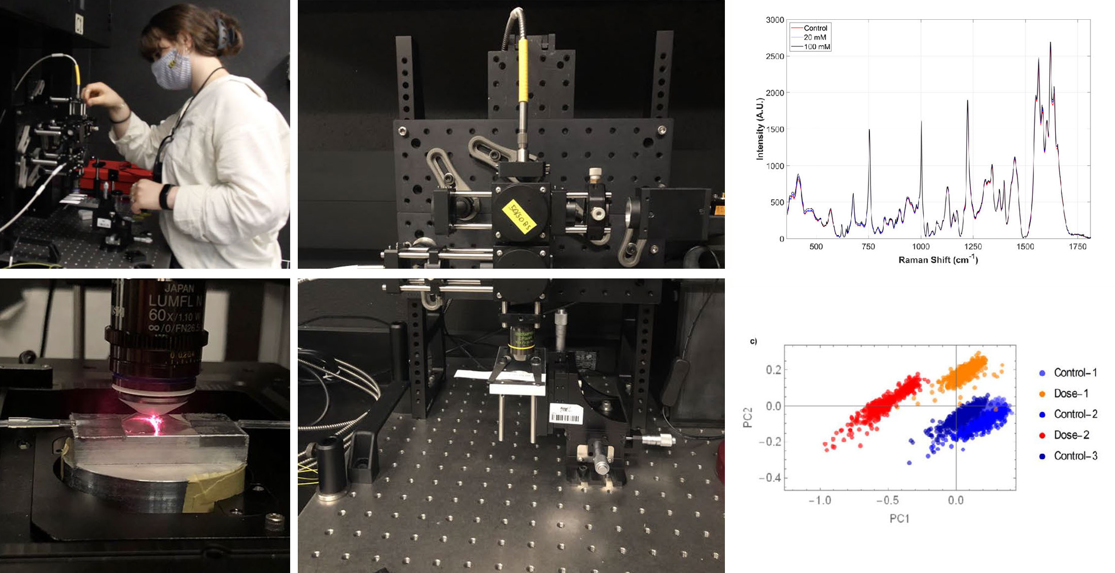
Using these techniques, we have successfully identified for example, ovarian cancer cells from healthy cells, chemo-sensitive vs resistant ovarian cancer cells, oxidized blood from control blood, and demonstrated novel through-the-surface detection of the composition of cartilage (e.g. for osteoarthritis) up to a depth of ~0.5 mm.
With our newly re-designed compact Raman flow-cell setup we are applying these techniques for distinguishing between control and irradiated blood, with collaborators at Health Canada. This has potential for biological dosimetry which is a key technique for retrospective radiation dosimetry that provides individual estimates of absorbed dose of ionizing radiation, applicable for use in a large scale radiological/nuclear event.
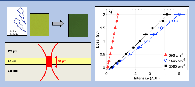
Ionizing radiation dosimetry based on high-resolution Raman spectroscopy readout of radiochromic films (RCFs)
Radiation therapy is used to treat about 50% of cancers world-wide; accurate measurement of the energy deposited is extremely important to optimize patient treatment. However techniques for ionizing radiation dosimetry using widely used chemical dosimeters such as radiochromic films (RCFs) are especially limited for micrometer-resolved dosimetry, at low and high doses of ionizing radiation. We recently reported a novel approach for high spatial resolution readout of RCFs using Raman spectroscopy. In contrast to previous work, we generated for the first time, dose calibration curves over an extended dose range from 0.03-50 Gy with uncertainty in the 5-8% range at 15-30 µm spatial resolution.
With the recently funded NFRF-E award, our team's goal is to develop a dosimetry system for cellular-level dose evaluation.
Coherent Raman, multimodal nonlinear optical imaging
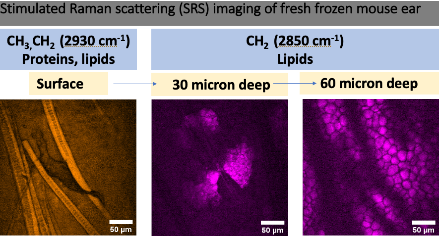
Coherent Raman imaging including Coherent Anti-Stokes Raman Scattering (CARS) and Stimulated Raman Scattering (SRS) belongs to a family of nonlinear optical (NLO) imaging techniques that provide rapid label-free, chemical imaging with high spatial resolution and inherent 3D sectioning capability. We have built a multimodal NLO imaging platform that is capable of SRS and CARS, combined with Second Harmonic Generation (SHG) and Two-Photon Excitation Fluorescence (TPEF) imaging. It employs a dual output laser with ~100 femtosecond pulses that are temporally broadened to a few picoseconds for CARS and SRS. The laser-scanning microscope is re-configurable and uses novel detection based on silicon photomultipliers (SiPMs) having important advantages over conventional photomultiplier tubes (PMTs). Our lab is a core member of the multi-disciplinary Tissue Engineering and Applied Materials (TEAM) Hub at Carleton.
Using this setup, we reported novel TPEF imaging-based metrics for quantifying the mitochondrial function and organization in immune cells that were treated with bacterial and viral load.
For a CIHR-funded project in collaboration with Prof. Fred Leblond and Dr. Dominique Trudel at the Centre de Recherche du Centre hospitalier de l'Université de Montréal (CRCHUM) in Montreal, we are currently developing SRS imaging-based techniques to identify an aggressive variant of prostate cancer in patient biopsies.
Development of compact Raman and NLOM systems
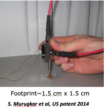
Sangeeta's experience in the photonics industry (JDS Uniphase) and her work as a Research Associate at University of Ottawa in Prof. Hanan Anis's lab, led to two photonics solutions that are significant for future clinical and industrial translation: i) the first portable miniaturized multimodal CARS microscope with a ~10,000x smaller footprint compared to a commercial microscope, and ii) a novel cost-effective (50% cheaper) excitation source for CARS imaging using a single femtosecond laser. At Carleton, we have built cost-effective (25% of the cost of commercial systems) platforms for Raman micro-spectroscopy and multimodal coherent Raman, NLO imaging. Our ultimate goal is to develop compact solutions for wider adoption in clinical and diagnostic lab settings.
Collaborators
The Centre hospitalier de l’Université de MontréalDr. Frederic Leblond and Dr. Dominique Trudel, Centre de recherche du Centre hospitalier de l'Université de Montréal | ||
 | University of OttawaDr. Barbara Vanderhyden, the Corinne Boyer Chair in Ovarian Cancer Research, University of Ottawa. | |
| University of OttawaDr. Hanan Anis and Dr. Robert Boyd, University of Ottawa. | |
 | University of TorontoDr. Brian Wilson, Department of Medical Biophysics, University of Toronto and the Princess Margaret Cancer Center, Toronto. | |
 | Health CanadaDr. Vinita Chauhan, Dr. Sami Qutob and Dr. Ruth Wilkins in the Consumer and Clinical Radiation Protection. | |
 | The Ottawa HospitalDr. Balazs Nyiri, Medical Physicist at the Ottawa Hospital. |
Funding
 |  |